
Prone OLIF and UBE after Four Previous Operations
19 August 2026 · Robotic
Presentation
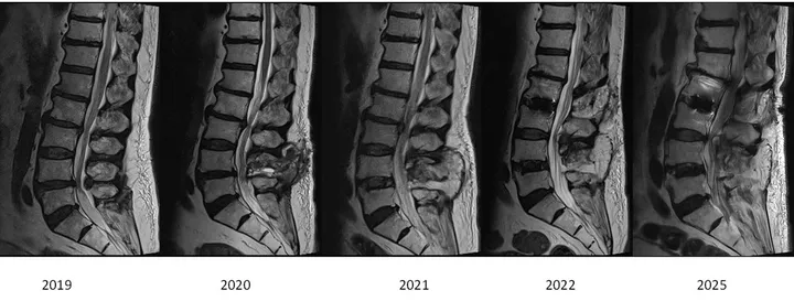
A man in his late 70s presented for a second opinion after more than six years of low back pain. He had already undergone four operations on his lumbar spine under another surgeon: a laminectomy at L4/5 in 2019, an XLIF with posterior fusion at L4/5 in 2020, a laminectomy at L2/3 in 2021, and an XLIF with posterior fusion at L2/3 in 2022.
By the time he was seen, his walking tolerance had fallen to 50–100 metres. He described altered sensation in both thighs and both shins, and there was no obvious weakness on examination.
He also reported bladder urgency. Any change in bladder function alongside nerve compression in the lower back is treated as a warning sign, and it moved the assessment forward without delay.
The challenge
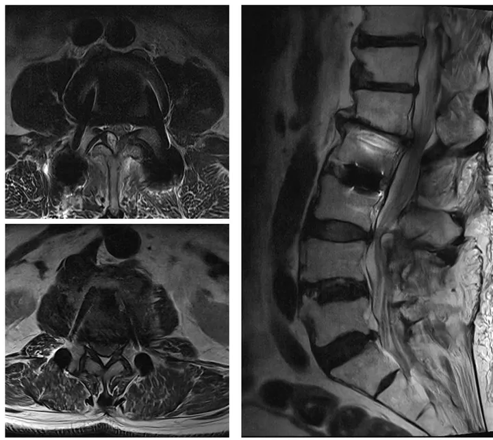
His imaging told a clear story when the scans were laid out in sequence. Between 2019 and 2025 the lumbar spine had been fused at L4/5 and again at L2/3, leaving a single mobile segment sandwiched between the two constructs. That remaining segment then carried the load of both fusions, and it progressively wore out — the disc collapsed, the canal narrowed, and the nerves passing through the level were compressed.
This is the situation that made the case difficult:
- Four previous operations meant extensive scar tissue in the midline and around the nerves, so re-opening the old posterior wound carried a real risk of injury to the dura and nerve roots
- Two sets of existing implants sat immediately above and below the level that now needed treatment
- The disc space was collapsed, so height had to be restored to reopen the space around the nerves
- His age and the length of his surgical history meant a shorter, gentler operation was preferable to a long open revision
One option was to perform the interbody fusion with him on his side and then turn and re-drape him to complete the posterior work. That repositioning step adds operative and anaesthetic time, so it was set aside in favour of a hybrid approach that could be completed in one position.
The approach
The plan was a prone OLIF (oblique lumbar interbody fusion) combined with UBE (unilateral biportal endoscopy) decompression, with robotic navigation, performed entirely with the patient lying prone.
- The disc space was reached through a small anterolateral incision along the anterior-to-psoas corridor, working in front of the psoas muscle and away from the scarred midline of the previous surgery.
- The collapsed disc was prepared with sequential, gentle distraction, and an interbody cage was placed to restore disc height. Restoring height alone reopens some of the space around the nerves — an indirect decompression.
- The nerves were then decompressed directly using UBE, a keyhole endoscopic technique performed through two small portals, which allowed the compressed structures to be seen and released under continuous irrigation and magnification.
- Fixation was added and connected to the implants already in place, so the previous constructs did not have to be removed.
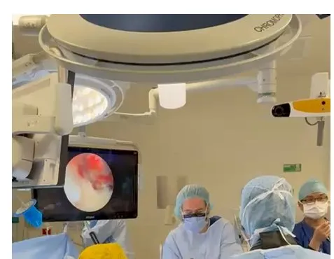
Because the anterior and posterior stages were both done prone, there was no need to reposition or re-drape partway through, which shortened the operative and anaesthetic time. Robotic navigation was used to plan and place the fixation, with instruments tracked on the navigation display rather than confirmed by repeated X-rays.
Imaging and result
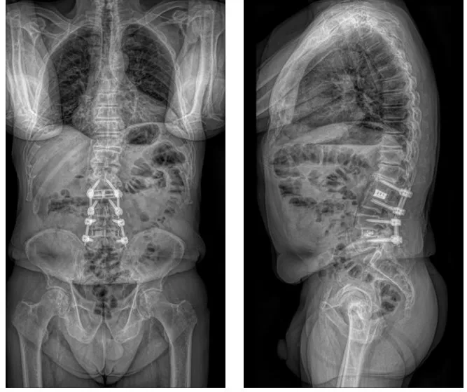
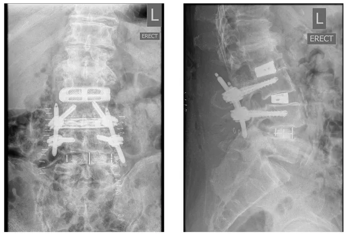
Intraoperative imaging confirmed the cage sitting in the disc space with height restored, and the new fixation joined to the earlier implants. Standing X-rays taken afterwards showed the completed construct under load. The figures below show the serial MRI sequence, the imaging before surgery, the theatre set-up and the final standing X-rays.
This case study is provided for education only and describes a single patient's course. Individual results vary — outcomes described here do not predict the results of treatment for any other patient.
We are here to listen. We understand your pain. Let us help.
Our professional team is at your disposal — from non-operative treatments through to advanced surgery.
Book a consultationor call 03 5925 6353 (Mornington) · 03 9989 3373 (Melbourne)