
Robotic-Assisted Prone OLIF and UBE at L4/5
19 August 2026 · Robotic
Presentation
A man in his 60s presented with long-standing low back pain that had recently changed character. Over the preceding three months he had developed progressive pain radiating into the left leg — the pattern of a compressed nerve root rather than back pain alone.
His walking tolerance had dropped to 50–100 metres before symptoms forced him to stop. On examination there were sensory changes in the left toes.
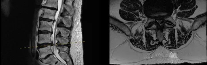
MRI showed degenerative change at L4/5 with narrowing of the canal at that level, matching the side and distribution of his leg symptoms.
The plan
The plan was a robotic-assisted prone OLIF at L4/5 combined with UBE decompression at the same level, in a single prone position.
The sequence mattered as much as the techniques. The principle was to correct first, then confirm and finish — restore the disc space and the alignment with the interbody stage, and only then use the endoscope to check the nerves directly and complete any decompression still required.
There is a practical reason for that order. Restoring disc height at a collapsed level reopens some of the space around the nerve roots on its own, so how much direct decompression is actually needed can only be judged accurately once the correction has been made. Doing the correction first avoids removing more bone and ligament than the case requires.
Before surgery, the fixation trajectories were planned on the patient’s imaging using the robotic platform. Planning at that stage means the intended screw path is defined in advance and the robotic arm then holds that trajectory in theatre, rather than the path being established freehand.
In theatre
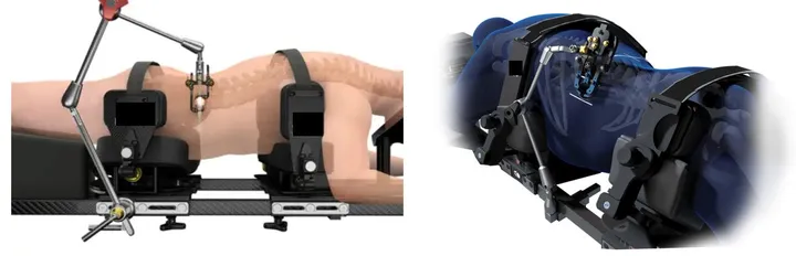
The patient was positioned prone on a positioning system with side padding, which supports the trunk while allowing the table to be tilted during the operation. That tilt is what makes the anterior approach workable with the patient face down.
- The disc space was reached along the anterior-to-psoas corridor through a small anterolateral incision, without entering the abdominal cavity.
- The disc was prepared and an interbody cage placed to restore height and correct the segment.
- Fixation was placed along the planned trajectories with robotic assistance.
- The nerves at L4/5 were then decompressed and inspected endoscopically through two small portals.
Because everything was done in one position, stages of the procedure could overlap rather than run strictly one after another, and there was no re-draping or repositioning step in the middle of the operation. In selected patients this reduces the amount of time spent under anaesthetic.
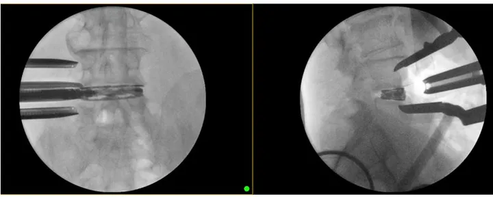
Fluoroscopy was used at intervals through the case — to confirm that disc height had been restored, to check the correction trajectory, and to confirm the final implant position before the patient left the room.
Result
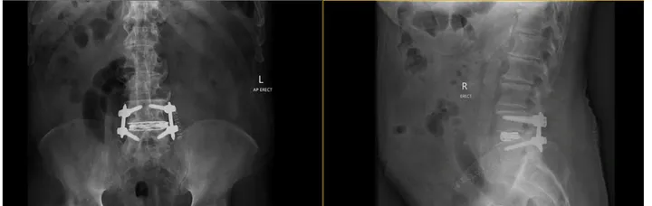
Standing X-rays after surgery showed the cage in the L4/5 disc space with the fixation in place. He recovered well, was mobilising on the first day after surgery, and was discharged home on the third day.
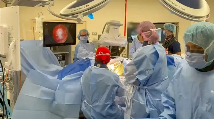
The figures below show the imaging before surgery, the prone positioning system, the theatre set-up, the intraoperative fluoroscopy and the final standing X-rays.
This case study is provided for education only and describes a single patient's course. Individual results vary — outcomes described here do not predict the results of treatment for any other patient.
We are here to listen. We understand your pain. Let us help.
Our professional team is at your disposal — from non-operative treatments through to advanced surgery.
Book a consultationor call 03 5925 6353 (Mornington) · 03 9989 3373 (Melbourne)